Sunday, March 20, 2011

Intermittent Fasting: Healthy or Harmful?


Healthy!  Resoundingly healthy.

Check out these sites for a comprehensive look at IF:


-Martin here has a great story.  He is a nutritional consultant, writer, and personal trainer.  Be sure and check out the testimonials and success stories from his clients.  Guaranteed, you'll be blown away.


- In my opinion, Mark has the premier site up now at MarksDailyApple when it comes to all aspects of Primal/Paleo eating and living.  He is incredibly well-spoken  and thorough in his posts and research.


Lose Weight Fast

-Brad has also got a good thing going with his book "Eat Stop Eat." He advocates including one or two 24 hour periods a week where you fast, both for fat loss and health benefits.  


- And be sure to check out Robb's work.  He has a great site and just wrote the book "The Paleo Solution: The Original Human Diet."  In it you can find some great advice and information on fasting.

Asymmetric Cell Division and Neural Stem Cells

This post will examine several things:
1.  What are stem cells? and what is ACD?
2.  What are the important features of ACD?
3.  Examples from model organisms.
4.  Diseases.

There are many different types of stem cells in our bodies.  Stem cells go on to produce specific cell populations in development.  For example, the ectoderm gives rise to neural stem cells which proliferate into neurons, skin, hair, and the mammory glands.

Stem cells have several properties.  First, they must be able to self-renew.  Second, they must possess a certain potency.  That is, they need to have the ability to divide to produce differentiated cells.  Totipotent stem cells can go on to produce every type of cell.  Pluripotent and multipotent stem cells are more limited in the cell populations that they can proliferate into, but they can still give rise to a large diversity of different cell populations.

Asymmetric cell division is the division of one cell that gives rise to two cells with different fates.  Normal cell division gives rise to two cells of equivalent fates.  Stem cells divide asymmetrically, giving rise to two distinct daughter cells, a copy of the original stem cell as well as another daughter with a non stem cell fate.
Centrosomes and the mitotic spindle play a key role in whether cells differentiate from the stem cells or not.

So how do stem cells divide?  Well, several factors come into play.  First, polarity cues provide the signal to divide or not.  There are both intrinsic and extrinsically-activated cues.  We also have asymmetrically localized determinants - cell polarity determinants and fate determinants.  Some common fate determinants are proteins (Numb being a key protein in determining neuronal fate), RNA, DNA, asymmetric phosphorylation, and organelles.  Mitotic spindle apparatus orientation also plays a big role.

Steps in ACD:
- Interphase: Setting up axis of cell/ Polarity cues
- Pro-metaphase: fate determinants segregated
- Metaphase: Mitotic spindle orients in cell
- Telophase: light coordination segregates components to different cells.

We can study stem cells in most of the common model organisms.  For example, big strides are being made in stem cell research in C. Elegans, yeast, C. Cerevisae, Drosophila, and mice (neural stem cells).

Neural stem cells function to produce neurons.  You can find them in both the PNS and the CNS.  They can include both pluripotent stem cells and multipotent progenitor cells.  And they can be found both during development and during adult homeostasis, interestingly enough.    

The Viral Me

Here's the link to GQ's article The Viral Me.  It is an honest look at the start-up scene and how it drives our new compulsion to take much of our lives into social media.

http://www.gq.com/news-politics/big-issues/201012/viral-me-silicon-valley-social-networking-devin-friedman

TEDx - Simon Sinek: How great leaders inspire action

It's not what you do; it's why you do it.


Apoptosis


Today's post is going to focus on cell death and pruning.  We are going to walk through the progressive and regressive processes that take place during brain development.

Some progressive events that take place during neuronal development include proliferation, axon guidance, and synapse formation.  Regressive events include apoptosis and axonal/dendritic/synaptic pruning.  Let's first take a look at the latter.

Both apoptosis and pruning are critical processes that sculpt the developing brain.

What are some factors that affect the survival of neurons during development?
Soluble factors include glia-derived and target-derived (retrograde) factors.  We also see afferent-derived (anterograde) factors, as well as hormones.

Types of cell death:
1.  Apoptosis: Leads to morphologic changes then cell death.  These changes include blebbing, loss of cell membrane asymmetry and attachment, cell shrinkage, nuclear fragmentation, chromatin condensation, and chromosomal DNA fragmentation. Unlike necrosis, apoptosis produces cell fragments called apoptotic bodies that surrounding cells are able to engulf and quickly remove before the contents of the cell can spill out onto surrounding cells and cause damage.*


2.  Necrosisthe premature death of cells and living tissue. Necrosis is caused by factors external to the cell or tissue, such as infection, toxins, or trauma. This is in contrast to apoptosis, which is a naturally occurring cause of cellular death. While apoptosis often provides beneficial effects to the organism, necrosis is almost always detrimental and can be fatal.*


Cell death machinery was first identified in C. Elegans.  With a total of only 959 cells, researchers found that 131 cells die during pruning, 26 of which are neurons.  Protein synthesis was crucial in determining whether a cell lived or died.  Anti-apoptotic proteins include Bcl-2 and Bcl-x.  Pro-apoptotic proteins include Bax, Caspase-9, Caspase-3, Apaf-1, and Cytochrome c.

Survival factors are NGFs, nerve growth factors, that aid in the survival of the cell.  Kind of self-explanatory.  Neurotrophin signaling activates anti-apoptotic genes and leads to survival.  From Wikipedia, "They [neurotrophins] belong to a class of growth factorssecreted proteins that are capable of signaling particular cells to survive, differentiate, or grow. Growth factors such as neurotrophins that promote the survival of neurons are known as neurotrophic factors. Neurotrophic factors are secreted by target tissue and act by preventing the associated neuron from initiating programmed cell death - thus allowing the neurons to survive. Neurotrophins also induce differentiation of progenitor cells, to form neurons."


Netrin-signaling is also anti-apoptotic.  


Sema/Plexin signaling is pro-apoptotic. 


As mentioned earlier, hormones also play a role in regulating cell death.  Metamorphosis in vertebrates and invertebrates is hormonally-regulated tissue deconstruction, associated with massive cell death and neuronal pruning.  Researchers have found evidence to suggest sexual dimorphism in circuits with regards to hormones.  One major study to show this involved bird mating songs.  They found that more neurons in the male birds were dedicated to mediating song acquisition compared to the females. Interestingly, they could induce masculinization of the female via injection of male-specific hormones.  


Neuronal Pruning: Pruning is a strategy often used to selectively remove exuberant neuronal branches and connections in the immature nervous system to ensure the proper formation of functional circuitry.  It can be small-scale and large-scale.  Small-scale pruning includes axosome shedding, lysosomal activity, activity-dependent pruning, and stochasticity.  You will see it in muscle cells and Purkinje neurons.  
Large-scale pruning is stereotyped/developmentally-regulated as well as activity-dependent.  


Take-away from this post:
1. Factors regulating axonal pruning:
- neural activity
- hormones
- guidance molecules - sema/plexins
- cohesin, ubiquitin-proteosome machinery, regulators of cytoskeletal dynamics, tx factors, caspases, RNA-binding proteins
2.  Regulation of regressive processes during neuronal development: Cross-talk between guidance mechanisms and cell death machinery
- guidance molecules as regulators of neuronal cell death
- cell death machinery (proteases, proteoltyic/lysosomal pathways) involved in axonal pruning.
- Pruning - local death of a neurite?  Cell death machinery is regulated by guidance cues.  


Ehhh, that was a long one.  
*Wikipedia

Synaptogenesis

...is the formation of synapses.  It involves axon guidance, topographic mapping, synapse formation, and synapse refinement.

Axon Guidance - As previously discussed on this blog in two parts, axon guidance involves long distance, intrinsic programs interacting with extrinsic cues.  See Axon Guidance - Part One and Axon Guidance - Part Two if you want to learn some more.

Topographic Mapping - medium distance, attractive and repellent gradients.  Topographic connections match behavior to the outside world.  Again, we can look at Sperry's Chemoaffinity Hypothesis...each target cell carries an id tag, the growing terminals of (RGC) cells have complementary tags, and the net effect is to permit RGC axons to seek out a specific location in the target region.

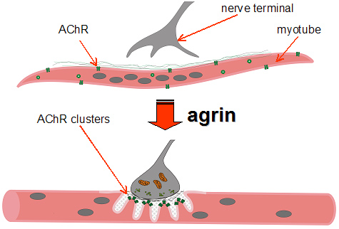
Synapse Formation - short distance, contact mediated.  Let's look at a specific molecule, Agrin.  Agrin is a proteoglycan that plays an integral role in the development of the NMJ during embryogenesis.  It also aids in the aggregation of ACh receptors during synaptogenesis.  Researchers found that pre- and post-synaptic elements at the NMJ are disrupted in agrin-deficient mice.  They found the exact same result in MuSK-deficient mice.  Agrin does not bind directly to MuSK but it does require MuSK to cluster AChRs.  Below is a figure detailing the multiple receptor signaling cascades that coordinate NMJ formation.

Synapse Refinement - near distance, activity-dependent.  Let's get into the specificity.  The brain favors molecular diversity; this requires synaptic elimination.  Connections that are not crucial or entirely necessarily will atrophy; crucial connections will strengthen.  If it's not required, it will be eliminated.  Synaptic refinement is a delicate and important process.  Again, it is obviously activity-dependent, and it occurs in the CNS as well.  Disrupting sensory activity perturbs refinement of synaptic connections.

And like most everything in the NS, when something goes wrong, we end up with disorders.  Angelman's, autism, and Fragile X are all implicated in dysregulation of synapse elimination.

Summary:

1.  Synaptogenesis involves multiple steps - axon guidance, topo mapping, synapse formation, synapse refinement.

2.  Maps are established by attractive and repulsive cues.

3.  Synapse formation involves both adhesive and inductive signaling events.

4.  Protein families involved in synaptogenesis include receptor tyrosine kinases (eg the EphRs and their ephrin ligands) and cell adhesion proteins (eg the Neuroligins and their neurexin ligands).

5.  Synapse specificity arises in part from molecular diversity (intrinsic) and then is refined by activity-dependent processes (extrinsic).

6.  Dysregulation of activity-dependent synapse refinement may contribute to neurodevelopmental disorders.  

Axon Guidance - Part Deux

What are the challenges for axon guidance in setting up the olfactory system?

An incredible example of precise axonal guidance is the axonal convergence of olfactory sensory neurons (OSNs) expressing a given odorant receptor (OR) onto spatially invariant glomeruli.  Evidence suggests that both guidance molecules and ORs play integral roles in the process.  Each OSN expresses a single type of OR.  In mice, the genome encodes around 1000 OR genes.  ORs are 7-transmembrane G-protein coupled receptors.  There are about 1 mil OSNs, each of which express 1 of the 1000 OR genes.  Therefore, on average, there are 1000 neurons expressing the same OR in the olfactory epithelium.  Neurons expressing the same OR are scattered in one of the zones in the epithelium.

Upon reaching the olfactory bulb, axons usually converge on one or two glomeruli.  There are around 1800 glomeruli, each of which has a topographically-fixed, invariant location in the olfactory bulb.  This means that somehow the axons sort, converge, and target the same glomerulus in the olfactory bulb with incredible precision.  Researchers have also found that swapping OR with B-adrenergic receptors also enables convergence of axons onto the glomeruli.  Pre-sorting of ORNs in the bulb depends on the level of cAMP generated by odorant (Sakano's model).  

TEDx - Yochai Benkler on the new open-source economics

Just watched this talk yesterday.  Great information on open-source.  Think Wikipedia and Red Hat...

http://www.ted.com/talks/yochai_benkler_on_the_new_open_source_economics.html

Roger Sperry - No Topsiders Here

In the 1940's, Roger Sperry did some interesting experiments on the frog visual system.  I'll discuss them in a minute, but first let's look at his Chemoaffinity Hypothesis.

"It seems a necessary conclusion...that the cells and fibers of the brain and cord must carry some kind of individual identification tags, presumably cytochemical in nature, by which they are distinguished one from another almost, in many regions, to the level of the single neuron..."  --Roger Sperry

In other words, each cell is specified by a distinct chemical label that signifies its address.  Axons then grow, as we will see, to match this address to the address in the target, forming complementary labels.

*"In the early 1940s, Roger Sperry performed a series of insightful experiments on the visual system of lower vertebrates that led him to draw two important conclusions: When optic fibers were severed, the regenerating fibers grew back to their original loci in the midbrain tectum to re-establish a topographical set of connections; and the re-establishment of these orderly connections underlay the orderly behavior of the animal. From these conclusions, he inferred that each optic fiber and each tectal neuron possessed cytochemical labels that uniquely denoted their neuronal type and position and that optic fibers could utilize these labels to selectively navigate to their matching target cell. This inference was subsequently formulated into a general explanation of how neurons form ordered interconnections during development and became known as the chemoaffinity hypothesis. The origins of this hypothesis, the controversies that surrounded it for several decades and its eventual acceptance, are discussed in this article."


Image
Fig. 4. Illustration of the optic nerve uncrossing surgery in frog and resulting behavior. In A, the normal optic chiasm is shown on the left wherein optic nerves cross to the contralateral side of the brain and the surgically uncrossed nerves are shown at the right wherein each optic nerve was deflected so as to regenerate to the ipsilateral side of the brain. In B, the mislocalization of flies by the frog with uncrossed optic nerves is shown by the position of the flies and the corresponding response indicated by the thick arrows. Taken from Scientific American article by Sperry.


*Taken from the abstract of Roger Sperry and his chemoaffinity hypothesis by Ronald L. Meyer

Axon Guidance

Back for some more neuroscience.  Today we are going to look at axon guidance, how it is achieved, and what determines whether guidance cues are short or long range, attractive or repulsive.

First off, what is axon guidance?  Axon guidance is the process by which developing neurons send out their axons to reach targets; the axons follow certain paths to the correct target.  We are going to look at how they manage to accurately reach their target.

An axon's growth cone, the motile tip of a growing axon, acts as a sensory vehicle, detecting cues in the extracellular environment and then reacting appropriately to those cues.  Guidance cues can either attract or repel the axon.  When the growth cone senses the guidance cues, it activates a series of intracellular cascades that ultimately lead to a change in the cytoskeletal structure of the neuron.

What kinds of molecular cues will an axon encounter?  There are several, and they can be grouped into two categories: Short vs. Long-range, and Attractive vs. Repulsive.

The axon will experience both attractive and repulsive cues many times over many different ranges.  Contact attraction and repulsion  is generally associated with short-range cues via surface proteins.  Chemoattraction and repulsion is more generally associated with a longer range.  Chemical signals are secreted and diffused within a distance of about 100-500um.

A good example of a class of repulsive guidance cues is the semaphorins.  They act as axon repellents by activating complexes of cell-surface receptors called plexins and neuropilins (class 3) and integrins (class 7).


Now we are going to examine an interesting and important case, crossing the midline.  One of the most crucial periods of human brain development involves axonal crossing of the midline, the forming of the corpus collosum, the nerve bundle connecting the two hemispheres of the brain, allowing for neural communication from one side to the other.

Kim Peek (the real-life "Rain Man"), a mega-savant, was born with agenesis of the corpus collosum and a missing anterior commisure.  His reading technique consisted of reading the left page with his left eye and the right page with his right eye.  He could therefore read two pages at a time, covering them at a rate of 8-10 seconds per page.  He could recall exact information when asked directly. So why couldn't one side of Kim's brain communicate with the other?  The decision to cross or not cross the midline is critical.  Let's take a closer look.

There are three very important steps in midline crossing:
1. Getting to the midline
2. Crossing it once
3. Moving on in the opposite hemisphere

Getting to the midline.  The commisural axons get to the midline via chemoattractant guidance cues. The floor plate plays an integral role in attracting the axons via emission of netrins, guidance proteins.  Sonic HeadgeHog (Shh) also acts to guide axons toward the floor plate. Netrins secreted by the floor plate cells function to bind the axon receptor DCC in a chemotactic manner. 

Crossing it once.  A study examining the ventral nerve cord of the fly (Goodman) found that axons cross the midline through the anterior commisure (AC) or posterior commisure (PC).  Wild-type fly axons crossed the midline once and left, continuing in their development in the opposite hemisphere.  However, researchers found 3 mutants that acted to disrupt the system: slit, robo, and comm.  Slit flies saw axons remain in the midline.  Roundabout (robo) flies saw axons cross the midline, only to recross back into the original hemisphere.  Commisureless (comm) flies saw no crossing of the midline.      

Moving on.   Once across, the axons generally have a decent rate of success in moving on.  Researchers have also come up with ways to switch on certain cues at different points in the process, enabling them to "choreograph" the sequence in different ways.


CODE Fun

Here's a fun problem to look at requiring Matlab programming. *

Many applications require us to know the temperature distribution in an object.  For example, this information is important for controlling the material properties such as hardness, when cooling an object formed from molten metal.  The following is a derivation of the temperature distribution in a flat rectangular metal plate.  The temperature on three sides is held constant at T1, and at T2 on the fourth side.  The temperature T(x,y) as a function of xy coordinates is given by

T(x,y) = (T2-T1)w(x,y)+T1

where

w(x,y)=(2/pi)*sum(((2/n)*sin((n*pi*x)/L)))*((sin((n*pi*y)/L)/(sin((n*pi*w)/L))));

The given data for the problem is as follows: T1=70*F, T2 = 200*F, w=L= 2ft.

Using a spacing of 0.2 for both x and y let's generate a mesh plot and contour plot of the temperature distribution.  Here is the code I wrote to try and solve the problem:


clear
clf
[x,y]=meshgrid(0:.2:2, 0:.2:2);
w=2;
L=2;
T1=70;
T2=200;
for n=1;
    wa=(2./pi).*((2./n).*sin((n.*pi.*x)./L)).*((sinh((n.*pi.*y)./L)./(sinh((n.*pi.*w)./L))));
end
for n=[1,3,5];
    wb=(2./pi).*((2./n).*sin((n.*pi.*x)./L)).*((sinh((n.*pi.*y)./L)./(sinh((n.*pi.*w)./L))));
end
for n=1:2:99
    wc=(2./pi).*((2./n).*sin((n.*pi.*x)./L)).*((sinh((n.*pi.*y)./L)./(sinh((n.*pi.*w)./L))));
end
%Fig 1
Ta=(T2-T1).*wa+T1;
subplot(1,2,1)
meshc(x,y,Ta)
subplot(1,2,2)
contour(x,y,Ta)
%Fig 2
Tb=(T2-T1).*wb+T1;
subplot(1,2,1)
meshc(x,y,Tb)
subplot(1,2,2)
contour(x,y,Tb)
%Fig 3
Tc=(T2-T1).*wc+T1;
subplot(1,2,1)
meshc(x,y,Tc)
subplot(1,2,2)
contour(x,y,Tc)


print -deps Palm5p56fig.eps


I tried to export my figure out of Matlab for you to include in here but couldn't quite figure it out.  Just run the code yourself and you'll get it though.

Enjoy!
*Adapted from Introduction to Matlab Programming for Engineers 7 by William Palm (pg 356)

Brainbow - The Multicolor Solution

What is Brainbow, and how can we use it? 


Brainbow is a new technique, developed in 2007 by Jeff W. Lichtman and Joshua R. Sanes, both professors of Molecular & Cellular Biology in the Department of Neurobiology at Harvard Medical School, allowing researchers to label individual neurons within a population with different colors.  


Brainbow leads to higher resolution than any previously-developed imaging methods, as well as quicker connectome mapping.  It helps to facilitate the surveying of quantitative and qualitative aspects of circuitry in diverse brain regions.  We can use Brainbow techniques to analyze connectional patterns in the brain over the lifespan of the organism.  Will this allow us to solve the mystery of the neurobiological underpinnings of healthy aging?  Only time will tell.  Solving this mystery, for example, would help us to more fully understand the vulnerabilities of an aging nervous system.  And on the other end of the spectrum, critical-period circuitry modification remains another mystery; brainbow techniques may open a door on them as well.  

The GAL4-UAS System

First developed by Andrea Brand and Norbert Perrimon in 1993, the GAL4-UAS system has since been considered a powerful biochemical method for studying genetic expression.  There are two parts to the system, the GAL4 gene (containing the GAL4 transcription-activating protein), and the UAS (Upstream Activating Sequence).  The UAS lies in the promoter region of the second chromosome.  The GAL4 protein binds to this region.  


For a bigger picture of a frequent application of the GAL4-UAS system, take this example (Wikipedia), "For example, suppose a scientist wants to visualize where a certain class of neurons extends to in the fly. He/she can then pick a fly from a GAL4 line that expresses GAL4 in the desired set of neurons, and cross it with a reporter line that express GFP. In the offspring, the desired subset of cells will make GAL4, and in these cells the GAL4 will bind to the UAS, and enable the production of GFP. So the desired subset of cells will now fluoresce green and can be followed with a microscope. Next suppose instead of looking at the cells, the experimenter wants to figure out what these cells do? One way is to express channelrhodopsin in each of these cells, by crossing the same GAL4 line with a channelrhodopsin reporter line. In the offspring the selected cells, and only those cells, will contain channelrhodopsin and can be triggered by a (bright) light. Now the scientist can trigger these particular cells at will, and perhaps find out what they do."

Model Organisms in Neuroscience

Neuroscientists primarily use six model organisms when doing research.  This post will examine these organisms and the advantages they provide scientists.

The model organisms are:
-Bacteria
-Yeast
-Arabidopsis
-C.Elegans
-Drosophila
-Mice

Why do we use these organisms, you might ask?  Some advantages of using model organisms are:
They have powerful genetics -- this means small genomes, rapid life cycles, transgenic ability, and they make it easy for us to map mutations.

A few specifics for the three most widely used organisms...
1.  C. Elegans - have very few cells (<1000 total -- 302 neurons), optically transparent, rapid development cycles, and canonical cell lineage. They are cheap and we know their entire connectome.  One of the more notable contributions of C.Elegans is the discovery of the Netrin pathway.  Other contributions include the discovery of the cell death genetic pathway, trigger for RNAi & systematic RNAi, the insulin pathway, first multicellular animal to have a completed genome, discovery of Par genes, and MicroRNA.

2.  Drosophila Melanogaster - have complex tissues and we can use sophisticated genetic techniques.  They are also cheap and have a short generation time.  We have sequenced their genome; drosophila has a rich history in the field of genetics.  They are a great model for studying the fundamental concepts in cell biology, developmental biology, and genetics.

3.  Mice - closest to humans, genetically.  We have sequenced their genome.  Since they have a more sophisticated nervous system, mice are generally used for studying immune, nervous, endocrine, and cardiovascular systems.


Neuron Doctrine

The neuron doctrine is the theory stating that the brain is made up of many tiny individual unit cells, neurons, that are interconnected via networks, yet still separate entities.

Single cell staining by dye impregnation was the first and most influential circuit-mapping strategy.  Employed by Santiago Ramon y Cajal (1852 - 1934), Golgi staining techniques (using potassium dichromate and silver nitrate) were used to stain neural networks.  This 'black reaction' staining technique stains just a few individual cells at a time, in their entirety, including all processes.  Cajal used it to stain networks and was subsequently able to discern the various types of neuronal networks in the brain, as well as describe their circuit organization.

We find several limitations with the single-cell staining techniques, however.  They cannot effectively do long-distance traces, nor are they effective when processes come within 0.25um of each other (cannot see it in the light microscope).  Researchers also have a difficult time distinguishing one cell from another, in some instances, if many elements in the same circuit are stained at once.

Mapping neural circuits is a central aim of modern neuroscience.  We need to develop methods of labeling/imaging neurons because the behavior of neural networks is critical to brain function and dysfunction and we need to see how the neurons are connected to better understand the networks.